MaxOne
Single-well · in-depthAnalyze one chip down to single-cell detail. Ideal for axon tracking, stimulation experiments, organoids and tissue slices.
Wells 1Recording 1,020 chNoise 2.2 µVrms
Discover innovative medical device solutions from microrobots to electrophysiology platforms.
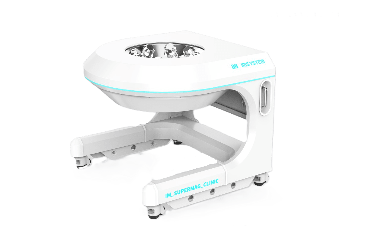
A next-generation magnetic microrobot system for precise interventional procedures, comprising a magnetic guidewire-microrobot and a precision magnetic field control system.

Comprised of a magnetic guidewire-microrobot and a precision magnetic field control system, the platform maintains compatibility with existing vascular intervention equipment to ensure clinical convenience.
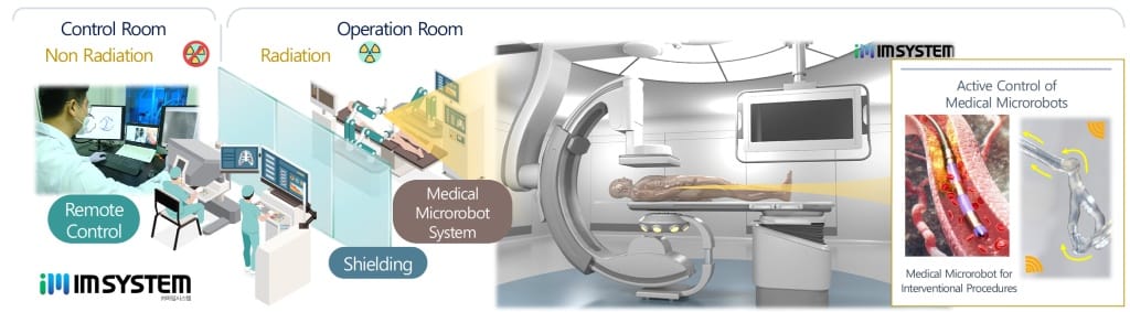
Using magnetic fields, the system precisely steers the intravascular magnetic guidewire-microrobot through complex vessel paths to reach the target site quickly and safely, performing treatment in conjunction with compatible tools such as catheters, stents, and balloons.
Reduces procedure time and improves success rates compared to conventional methods. Remote operation minimizes radiation exposure for medical staff, while also reducing fatigue from lead apron use and the risk of infectious disease transmission.
The system consists of remote control, electromagnetic coils, compatibility with conventional equipment, and a magnetically steerable guidewire-microrobot.

Remote operation means less radiation exposure for the medical crew.
Generates a 3D magnetic field to control the direction of the magnetic guidewire-microrobot.
Compatible with X-ray imaging systems and the patient bed for clinical convenience.
Steered precisely by the magnetic field through complex vessels to reach the target site.
The medical crew operates the system remotely to minimize radiation exposure, while the magnetic field steers the guidewire-microrobot inside the vessel.
Magnetic guidewire-microrobot tests in vascular phantoms and a preclinical coronary intervention in swine, using the magnetic field control system.

Future development of biomedical microrobot systems will cover a wider range of treatments for various diseases.

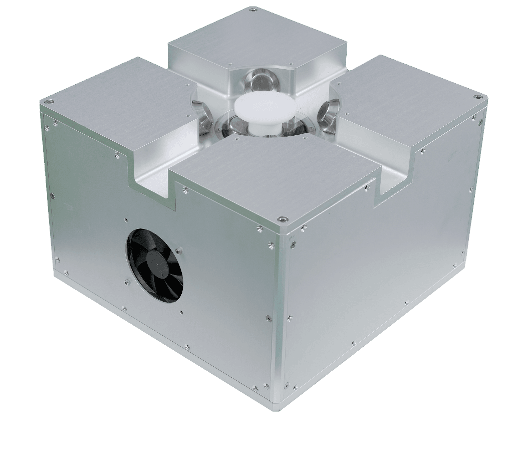

Magnetic field system for precise control of magnetic objects in lab-scale in vitro and preclinical research
A 3D magnetic field precision control system for lab-scale research. By manipulating the position and orientation of magnetic objects, it can be used for in vitro experiments, preclinical studies and other magnetic research.
Supports constant, rotating and gradient fields, programmed sequential control, real-time current and temperature monitoring, and 5-DOF magnetic field control.
Monitors coil current and temperature in real time
3-axis translation and 2-axis rotation
Consists of the electromagnet coil, the current control module (ECB) and the user interface software.
Magnetic Field Generation
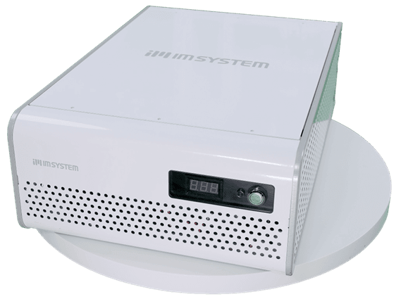
Current Control & Monitoring
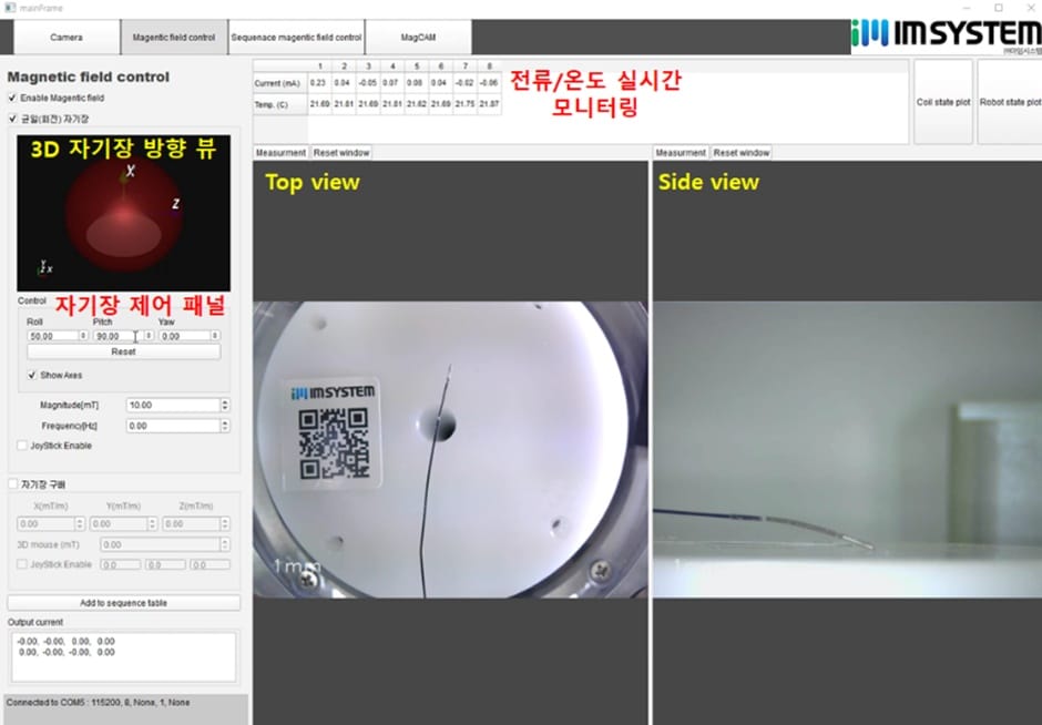
Magnetic Field Control & Visualization
Controls the orientation and position of magnetic objects, microrobots, magnetic particles and magnetic cells with magnetic fields.


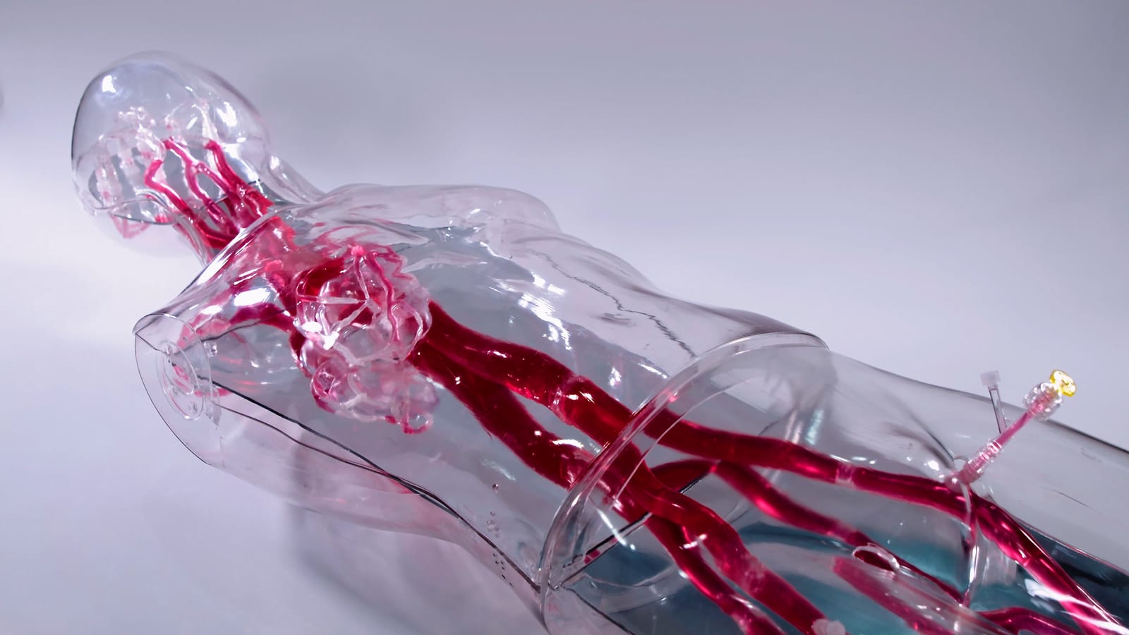
A full-body 3D vascular simulator that precisely replicates the major vessels from head to lower limbs based on real anatomical diameters. Internal lubrication coating provides a realistic procedural feel, making it ideal for multi-site vascular intervention training, medical device validation and education.
Using precision-matched transparent silicone with internal lubrication coating, VasculiX enables realistic vascular intervention training and medical device validation in an X-ray compatible environment.
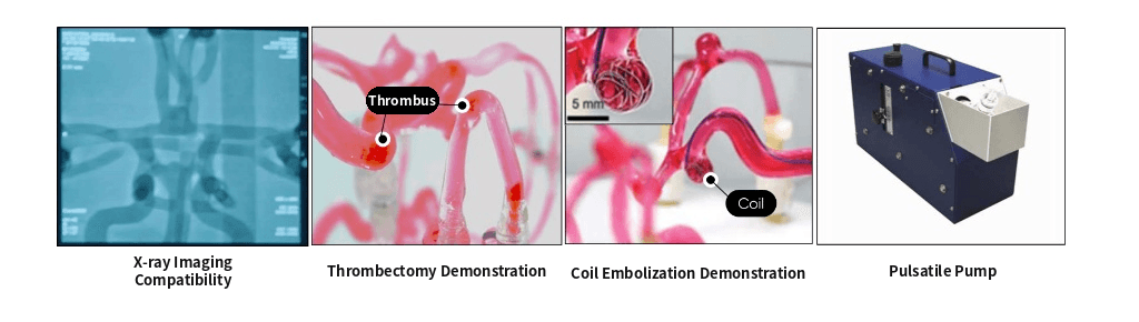
A specialized internal coating technology replicates the friction coefficient (0.07–0.15) of real vessel walls. Catheter and guidewire manipulation feels identical to actual procedures, maximizing training fidelity. Friction characteristics can be customized per user request.
Silicone is radiolucent and barely blocks X-rays, allowing real-time visualization of intravascular device movement under fluoroscopy (C-arm, DSA). Supports contrast injection experiments and stent/coil placement verification under authentic interventional conditions.
Custom vascular models replicate conditions such as cerebral aneurysms, atherosclerosis, and thrombus, enabling live demonstration of coil embolization, thrombectomy, and other interventional procedures. Connection to a pulsatile pump reproduces realistic cardiac-cycle blood flow.
Patient-specific CT/MRI data can be used to fabricate custom vascular models that precisely replicate individual anatomy. Standard models optimized for medical device R&D and bench-test validation are also available, with freely configurable vessel diameter, curvature, and branching geometry.
Comparison of procedural conditions with and without coating

A 3D cerebrovascular simulator precisely replicating real anatomical diameters — from Common Carotids to the Circle of Willis and cerebral aneurysms (A·B·C). Vessel wall thickness is fully adjustable, and internal lubrication coating provides a tactile feel identical to real procedures. Optimized for neuro-interventional training and medical device demonstration and research.
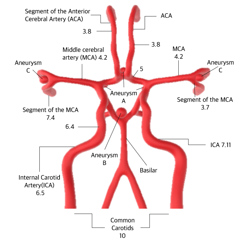
Common Carotids 10mm · ICA 6.5~7.11mm · MCA 4.2mm · ACA 3.8mm
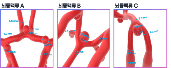
Aneurysm A (Circle of Willis bifurcation) · Aneurysm B (ICA segment) · Aneurysm C (MCA distal)
VasculiX_Neuro precisely replicates major cerebrovascular structures from the Common Carotids to the full Circle of Willis, based on real anatomical diameter data. Pathological features including cerebral aneurysms (Aneurysm A·B·C) are also reproduced, providing an interventional environment identical to clinical conditions.
Common Carotids
Diameter 10mm — Entry point of the cerebrovascular model. Bifurcates into left and right internal carotid arteries
Internal Carotid Artery (ICA)
Diameter 6.5~7.11mm — Major artery entering the cranium. Includes Aneurysm B lesion segment
Basilar Artery
Diameter 6.4mm — Posterior component of the Circle of Willis. Includes Aneurysm A (5mm) at the bifurcation
Middle Cerebral Artery (MCA)
Diameter 4.2mm — Supplies blood to the lateral cerebral cortex. Includes MCA segments (3.7~7.4mm) and Aneurysm C
Anterior Cerebral Artery (ACA)
Diameter 3.8mm — Terminal segment artery supplying the medial cerebral surface and frontal lobe
Using precision-replicated cerebrovascular structures in transparent silicone with internal lubrication coating and acrylic packaging, VasculiX_Neuro provides an optimized environment for neuro-interventional procedure training and medical device validation.
A specialized internal coating technology replicates the friction coefficient (0.07–0.15) of real vessel walls. Catheter and guidewire manipulation feels identical to actual procedures, maximizing training fidelity. Friction characteristics can be customized per user request.
Silicone is radiolucent and barely blocks X-rays, allowing real-time visualization of intravascular device movement under fluoroscopy (C-arm, DSA). Supports contrast injection experiments and stent/coil placement verification under authentic interventional conditions.
Custom vascular models replicate conditions such as cerebral aneurysms, atherosclerosis, and thrombus, enabling live demonstration of coil embolization, thrombectomy, and other interventional procedures. Connection to a pulsatile pump reproduces realistic cardiac-cycle blood flow.
Patient-specific CT/MRI data can be used to fabricate custom vascular models that precisely replicate individual anatomy. Standard models optimized for medical device R&D and bench-test validation are also available, with freely configurable vessel diameter, curvature, and branching geometry.
Comparison of procedural conditions with and without coating
Two options depending on the internal case configuration
Case interior is filled with air; the vessel simulator stands alone. Lightweight and optimized for structural visualization. This is the standard specification.
Case interior is filled with silicone to support the vessel simulator on all sides. Mimics surrounding tissue mechanical properties for a more realistic procedural environment.

A 3D cardiovascular simulator that precisely replicates real anatomical diameters — from the aortic root, where the coronary arteries originate, through the aortic arch, carotid and subclavian arteries, to the descending aorta. Vessel wall thickness is adjustable, and internal lubrication coating provides a realistic procedural feel. Optimized for percutaneous coronary intervention (PCI) training and medical device demonstration and research.
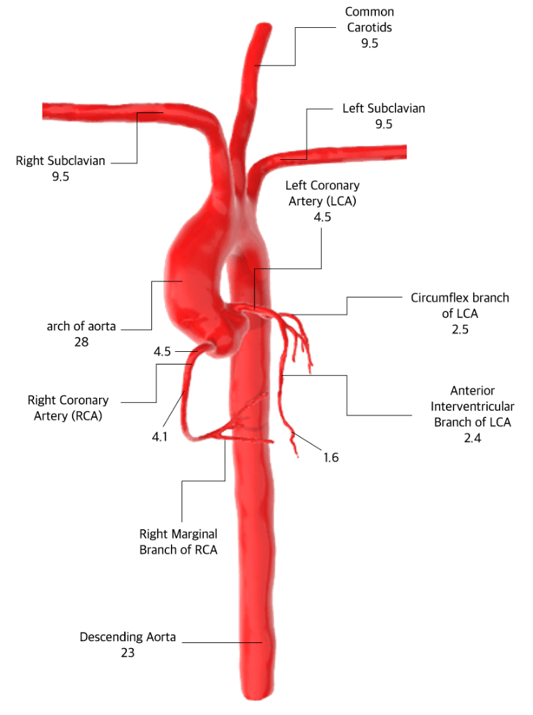
Arch of Aorta 28mm · Left Coronary Artery (LCA) 4.5mm · Right Coronary Artery (RCA) 4.1mm · Descending Aorta 23mm
VasculiX_Cardio precisely replicates major cardiovascular structures — including the aorta and coronary arteries — based on real anatomical diameter data. The geometry and diameter of each vascular branch are designed according to clinical literature standards.
Common Carotids
Diameter 9.5mm — Major cervical artery supplying blood to the brain
Left / Right Subclavian Artery
Diameter 9.5mm — Artery supplying blood to the arms and shoulders
Arch of Aorta
Diameter 28mm — Arch segment of the largest artery exiting the heart
Left Coronary Artery (LCA)
Diameter 4.5mm — Coronary artery supplying the left side of the heart. Branches into the Left Circumflex (LCx, 2.5mm) and Left Anterior Descending (LAD, 2.4mm) arteries
Right Coronary Artery (RCA)
Diameter 4.1mm — Coronary artery supplying the right side of the heart. Branches into Right Marginal Branch (1.6mm)
Descending Aorta
Diameter 23mm — Descending segment of the aorta running inferiorly from the aortic arch toward the abdomen
Using precision-replicated coronary and aortic structures in transparent silicone with internal lubrication coating and acrylic packaging, VasculiX_Cardio provides an optimized environment for cardiovascular intervention training and medical device validation.
A specialized internal coating technology replicates the friction coefficient (0.07–0.15) of real vessel walls. Catheter and guidewire manipulation feels identical to actual cardiovascular procedures, maximizing training fidelity. Friction characteristics can be customized per user request.
Silicone is radiolucent and barely blocks X-rays, allowing real-time visualization of intravascular device movement under fluoroscopy (C-arm, DSA). Supports contrast injection and stent/balloon placement verification under authentic cardiovascular intervention conditions.
Custom vascular models replicate cardiovascular conditions such as coronary stenosis, atherosclerosis, and aortic stenosis, enabling live demonstration of coronary stenting, balloon angioplasty, and other cardiovascular interventional procedures. Connection to a pulsatile pump reproduces realistic cardiac-cycle blood flow.
Patient-specific CT/MRI data can be used to fabricate custom models that precisely replicate coronary and aortic anatomy. Standard models optimized for cardiovascular medical device R&D and bench-test validation are also available, with freely configurable vessel diameter, curvature, and branching geometry.
Comparison of procedural conditions with and without coating
Two options depending on the internal case configuration
Case interior is filled with air; the vessel simulator stands alone. Lightweight and optimized for structural visualization. This is the standard specification.
Case interior is filled with silicone to support the vessel simulator on all sides. Mimics surrounding tissue mechanical properties for a more realistic procedural environment.
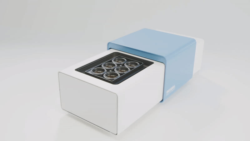
MaxWell Biosystems HD-MEA systems that record extracellular electrical activity of cells on a CMOS chip with 26,400 electrodes. MaxOne is a single-well system; MaxTwo is a multi-well system with 6- or 24-well plates.
Analyze one chip down to single-cell detail. Ideal for axon tracking, stimulation experiments, organoids and tissue slices.
Record many conditions at once on 6-well and 24-well plates. Ideal for dose comparisons, toxicity assessment and large cell-line screens.
MaxOne · MaxTwo brochure — specifications, case studies and chip types in one PDF.
Download Brochure (PDF)Key applications, what each one measures, and which system fits.
 Measured data
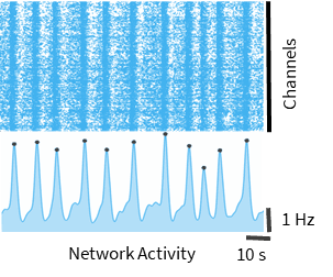
Measure firing and synchrony in cultured neuronal populations to see how the network communicates.
 Measured data
Record the same organoid repeatedly over weeks to track maturation and disease-model differences non-invasively.
Map how a neuron's action potential travels along its axon at µm resolution to measure conduction velocity and branching.
Measure beating and signal propagation in iPSC-derived cardiomyocytes and check rhythm changes under drugs.
Dose multiple wells at different concentrations and record them together to compare neuro- and cardiotoxicity in one run.
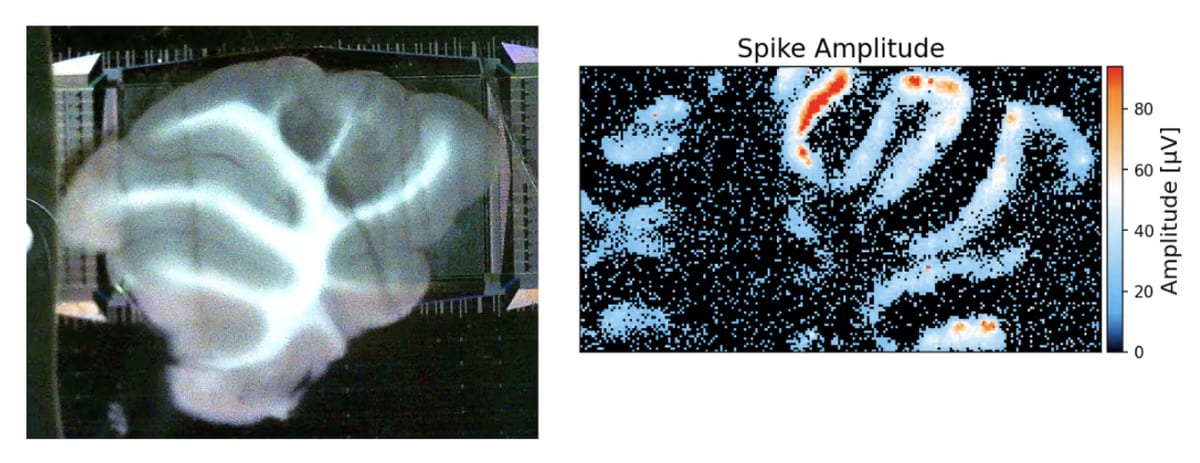 Measured data
Place acute brain slices or retina on the chip to map, in space, how tissue circuits respond to stimulation.
The applications above combine these four assays. All four run on both MaxOne and MaxTwo in MaxLab Live software.
Scans all 26,400 electrodes to find where signals occur.
Records selected electrodes together to analyze bursts and synchrony.
Maps how one neuron's signal travels along its axon.
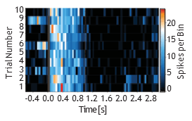
Stimulate cells at chosen electrodes and record the response — up to 32 electrodes at once.
| MaxOne | MaxTwo | |
|---|---|---|
| Best for | One sample in depth · down to individual cells | Many samples at once · comparing conditions |
| Format | 1 well (one chip) | 6-well / 24-well plate |
| Electrodes | 26,400 per chip (well) · 17.5 µm pitch · 20 kHz (both systems) | |
| Typical uses | Neuronal networks · organoids · axons · brain slices & retina | Drug screening · toxicity · cardiomyocytes · axon comparison across wells |
Planar Standard MEA
Planar Large MEA
| Feature | PSM | PLM |
|---|---|---|
| Active sensing area | 3.85 × 2.10 mm² | |
| Total number of electrodes | 26,400 | |
| Electrode density | 3,265 electrodes/mm² | |
| Electrode center-to-center distance | 17.5 µm | |
| Electrode size | 11.5 × 11.5 µm² | |
| Electrode material | PEDOT / Platinum | |
| Surface topography | <0.6 µm | <0.3 µm |
| Sampling rate | 20 kHz/channel | |
| Number of recording channels | 1,020 | |
| Typical noise per chip | 2.2 µVrms | |
| Electrical stimulation | ✓ Yes | |
| Electronic ID | ✓ Yes | |
| Sterilization Treatment | ✓ Yes | |
Tell us about your samples and goals and we'll recommend a setup and assays. Demo measurements are available.
Interested in medical microrobot technology?
Consult with IMsystem experts.